超声成像_人工智能如何帮助转变医学超声成像
超声成像
介绍 (Introduction)
Diagnostic ultrasound is a popular imaging method used for a variety of screening and diagnostic procedures such as pregnancy monitoring, thyroid screening, blood flow evaluation or breast cancer detection. Most of these examinations are performed by highly-trained clinicians specializing in these areas. Since the ultrasound machine is a highly complex instrument, it requires months or even years of experience to acquire the proper diagnostic skillset. Ultrasound examinations may take a long time (up to 1 hour) even for skilled professionals.
诊断超声是一种流行的成像方法,可用于多种筛查和诊断程序,例如妊娠监测,甲状腺筛查,血流评估或乳腺癌检测。 这些检查大多数由专门研究这些领域的训练有素的临床医生进行。 由于超声仪是一种高度复杂的仪器,因此需要数月甚至数年的经验才能获得适当的诊断技能。 即使是熟练的专业人员,超声波检查也可能需要很长时间(最多1个小时)。
In many medical fields, ultrasound is still underutilized. We can identify the following reasons for this development:
在许多医学领域,超声技术仍未得到充分利用。 我们可以确定导致这种发展的以下原因:
Lack of clinician experience, making it easier to refer the patient to an expensive CT or MR procedure.
缺乏临床医生经验 ,因此更容易将患者转介至昂贵的CT或MR手术。
Long examination time, leading to reduced patient throughput. This is especially noticeable in smaller practice clinics and point-of-care medicine, where other diagnostic procedures may be preferable to imaging due to time constraints.
较长的检查时间 ,导致患者吞吐量下降。 这在小型诊所和即时医疗中尤其明显,由于时间限制,其他诊断程序可能比成像更可取。
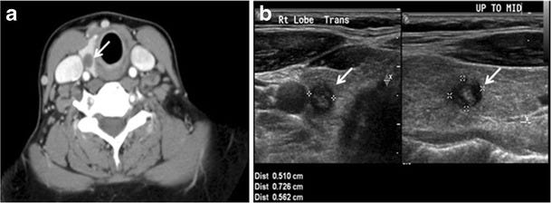
Ultrasound images may be harder to read compared to X-ray or CT scans due to the inherent noise (speckle) and reduced contrast.
由于固有噪声(斑点)和对比度降低,与X射线或CT扫描相比, 超声图像可能更难以读取 。
 Insights into Imaging 成像洞察 7 (4): 601–617. 7 (4):601–617。 DOI:DOI : 10.1007/s13244–016–0506–5. 10.1007 / s13244–016–0506–5 。 ISSN ISSN 1869–4101.1869–4101 。
 As an example of underutilization, consider point-of-care (bedside) medicine. Procedures in this area are often performed by less-skilled clinicians as well as nurses. Therefore, specialized ultrasound training is scarce. Moreover, since time is a crucial variable in this field (point-of-care medicine often deals with acute issues), examinations must be kept as short as possible. Imaging usually isn’t a great tool when it comes to speed.
作为未充分利用的一个例子,考虑即时医疗(床边) 。 该领域的手术通常由技术欠佳的临床医生以及护士来执行。 因此,缺乏专门的超声训练。 而且,由于时间是该领域的关键变量(即时医疗经常处理急性问题),因此必须尽可能缩短检查时间。 就速度而言,成像通常不是一个很好的工具。
Recently, AI algorithms claimed very good performance in diagnosing various diseases from medical images, sometimes even surpassing human-level performance. Although most of these studies concentrate on CT or MRI imaging, analyzing ultrasound images using AI has also been studied quite extensively. On the other hand, it is hard to imagine that AI will completely replace humans for the diagnostic procedure anytime soon. This is even more true with ultrasound, since the operator has to manually scan the patient. Instead, exploring how AI might simplify life for the clinicians while keeping the ultimate responsibility for the diagnosis with the human expert may be more valuable in the short term.
最近, 人工智能算法声称在从医学图像诊断各种疾病方面具有非常好的性能 ,有时甚至超过了人类水平的性能。 尽管这些研究大多数集中在CT或MRI成像上,但使用AI分析超声图像也得到了广泛的研究。 另一方面, 很难想象AI将在不久的将来完全取代人类来进行诊断程序。 对于超声波,情况更是如此,因为操作员必须手动扫描患者。 相反,在短期内,探索AI如何简化临床医生的生活,同时与人类专家保持诊断的最终责任可能更有价值。
In this article, I will first explain how an ultrasound exam usually works. In the second part, I will focus on where AI might jump in to improve the quality and efficiency of the procedure. Finally, I will try and make a few predictions about how fast the ultrasound AI revolution might happen.
在本文中,我将首先解释超声检查通常如何工作。 在第二部分中,我将重点介绍AI可能会进入的地方,以提高过程的质量和效率。 最后,我将尝试就超声波AI革命发生的速度做出一些预测。
超声检查的解剖 (Anatomy of an Ultrasound Examination)
Essentially, an ultrasound examination consists of the following parts:
本质上,超声波检查包括以下部分:
- The patient enters the scanning room. 病人进入扫描室。
- The doctor prepares the ultrasound machine and adjusts the settings for the upcoming exam (ultrasound probe, scanning preset). 医生会为超声仪做准备,并为即将进行的检查(超声波探头,预设扫描)调整设置。
- Additional patient-specific adjustments may be necessary. 可能需要其他针对患者的调整。
- The doctor finds the corresponding anatomy(-ies) by moving the probe to the correct location(s). 医生通过将探头移动到正确的位置来找到相应的解剖结构。
- The doctor annotates important landmarks and performs measurements. 医生注释重要的地标并进行测量。
- The doctor saves screenshots and moves on to the next anatomy or ends the live exam. 医生将保存屏幕截图,然后继续进行下一个解剖或结束实时检查。
- The doctor reviews the screenshots and finalizes the diagnosis. 医生检查屏幕截图并完成诊断。
Since these steps have to be performed in nearly every exam and they are mostly manual, there is a lot of room for workflow improvement. Furthermore, most of the steps above consist of multiple substeps that have to be performed repeatedly (e.g., making a measurement consists of selecting the measurement tool in the user interface, placing corresponding calipers and naming the measurement).
由于这些步骤几乎在每次检查中都必须执行,并且大多数都是手动操作,因此有很大的改进空间 。 此外,上面的大多数步骤都包含多个子步骤,这些子步骤必须重复执行(例如,进行测量包括在用户界面中选择测量工具,放置相应的卡尺并命名测量)。
Some steps, such as finding the correct view, can be performed very quickly by experienced users, while less experienced users may struggle. This is also influenced by the image quality of the instrument. Since better trained and more experienced users perform the same examinations as the less trained ones, the quality of the examination and therefore the diagnosis may vary wildly.
有经验的用户可以非常快速地执行某些步骤,例如查找正确的视图,而经验不足的用户可能会遇到困难。 这也受到仪器图像质量的影响。 由于训练有素和经验更丰富的用户执行的检查与训练有素的用户相同,因此检查的质量以及诊断可能会发生巨大差异。
AI如何提供帮助 (How AI Might Help)
Where in the ultrasound examination may we insert a bit of AI magic?
在超声波检查中,我们可以在哪里插入一些AI魔术?
Let’s look at the list we assembled in the previous section and dissect it:
让我们看一下我们在上一节中汇编的列表并进行剖析:
- When preparing for the exam and selecting the ultrasound probe and preset, there isn’t too much you can improve, so we will leave that step untouched. 在准备检查并选择超声探头和预设时,您没有太多可以改进的地方,因此我们将保持这一步骤不变。
Additional patient-specific adjustments can be made using AI though. Although this is not yet widespread in ultrasound, the company ContextVision already offers AI-based image enhancement for radiography (https://www.contextvision.com/products/radiography/).
不过, 可以使用AI进行其他针对患者的调整 。 尽管这在超声领域尚未广泛应用,但是ContextVision公司已经提供了基于AI的射线照相图像增强功能( https://www.contextvision.com/products/radiography/ )。
Finding the corresponding anatomy is one of the most difficult tasks for inexperienced operators. Unfortunately, it is hard to completely automate this step, since it involves moving the probe itself. However, AI can help with the navigation: e.g., the FDA-approved tool Caption Guidance can help the user acquire standard views of the heart. A similar solution is developed by the company Intelligent Ultrasound. An additional opportunity is presented through the use of 3-D ultrasound: although the image quality of sections of the acquired 3-D volume is usually inferior to that of a 2-D ultrasound, it is often sufficient for accurate diagnosis. A tool developed by GE Healthcare for AI-based plane recognition in the fetal brain called SonoCNS provides this kind of functionality.
对于没有经验的操作员来说,找到相应的解剖结构是最困难的任务之一。 不幸的是,由于此步骤涉及移动探针本身,因此很难完全自动执行此步骤。 但是, AI可以帮助导航 :例如,FDA批准的工具Caption Guidance可以帮助用户获取心脏的标准视图。 Intelligent Ultrasound公司也开发了类似的解决方案。 通过使用3-D超声提供了另一个机会:尽管获取的3-D体积的各个部分的图像质量通常不如2-D超声的图像质量,但对于准确的诊断通常就足够了。 GE Healthcare开发的一种用于胎儿大脑中基于AI的平面识别的工具SonoCNS提供了这种功能。
 GE HealthcareGE Healthcare
 Making annotations and measurements may also be simplified greatly (and sometimes completely automated) by AI. After finding the corresponding planes, SonoCNS can automatically perform some standard measurements of the fetal brain. More complex measurements around cardiac screening are also being automated: Ultromics offers automated cardiac analysis in their EchoGo Core software (FDA-approved). EchoGo is able to calculate the left ventricular ejection fraction, left ventricular volumes and even cardiac strain. DiA’s LVivo RV provides fully automated analysis of the heart’s right ventricle (also relevant for COVID-19 patients) while their LVivo Bladder provides automated bladder volume measurements (also FDA-approved). PIUR Imaging is the first to provide AI-assisted analysis of carotid plaque, including plaque volume, volumetric stenosis, 3D grayscale median and grayscale median distribution. The results are summarized in a report as shown below.
AI也可以极大地简化注释和测量(有时是完全自动化)。 找到相应的平面后,SonoCNS可以自动执行胎脑的一些标准测量。 围绕心脏筛查进行的更复杂的测量也已实现自动化:Ultromics在其EchoGo Core软件 (FDA批准)中提供了自动化心脏分析。 EchoGo能够计算出左心室射血分数,左心室容积甚至心脏劳损。 DiA的LVivo RV提供了心脏右心室的全自动分析(也与COVID-19患者相关),而他们的LVivo膀胱提供了膀胱自动测量(也经过FDA批准)。 PIUR Imaging是第一个提供AI辅助分析颈动脉斑块的方法,包括斑块体积,体积狭窄,3D灰度中位数和灰度中位数分布。 结果汇总在报告中,如下所示。
 PIUR ImagingPIUR Imaging
 AI can also automate screenshot-taking: screenshots can be saved as soon as the (automated) measurements were performed. In addition, the AI may guide the clinician through the entire exam — since it is familiar with the anatomies being scanned, it knows what remains to be examined. As soon as all the standard views were acquired, the software can automatically notify the clinician that the exam is over.
AI也可以自动进行屏幕截图 :执行(自动)测量后即可保存屏幕截图。 另外,AI可以指导临床医生完成整个考试-由于它熟悉所扫描的解剖结构,因此它知道需要检查的内容。 一旦获取了所有标准视图,该软件即可自动通知临床医生检查已结束。
未来什么时候到来? (When Will the Future Arrive?)
As already mentioned, the first AI-supported applications are on the market and they are getting more and more widespread as new clinicians are increasingly relying on them. All leading manufacturers of ultrasound machines as well as several startups have caught up to this trend and invest heavily in this direction. Medical cloud solutions, especially in North America and Asia, are on the rise. Another argument in favor of AI is the rising demand on patient throughput. This will only be manageable with rising levels of automation.
如前所述,第一个支持AI的应用程序已投放市场,并且随着新的临床医生越来越依赖它们,它们也越来越广泛。 所有领先的超声机制造商以及多家初创公司都已顺应这一趋势,并在此方向上进行了大量投资。 医疗云解决方案,特别是在北美和亚洲,正在上升。 支持AI的另一个论点是对患者通量的需求不断增长。 只有随着自动化水平的提高,这才可以管理。
On the other hand, clinicians are ultimately still responsible for the decisions they make with respect to the individual patients. Therefore they are understandably skeptical about the use of AI in their domain. This is especially true for highly-skilled doctors with many years of experience under their belt. Recent research on adversarial attacks on neural networks really should not make the doctors or the patients feel any more comfortable. I think this will change as the number of AI applications will rise and more and more clinicians will start taking advantage of the technological improvements. This will in turn provide more feedback to the developers, feeding the improvement cycle.
另一方面,临床医生最终仍要对他们就个别患者做出的决定负责。 因此,他们对在自己的领域使用AI表示怀疑,这是可以理解的。 对于拥有多年经验的高技能医生来说尤其如此。 最近对神经网络进行对抗性攻击的研究确实不应使医生或患者感到更舒服。 我认为随着AI应用程序数量的增加以及越来越多的临床医生将开始利用技术进步,这种情况将会改变。 反过来,这将为开发人员提供更多反馈,从而满足了改进周期。
My prediction is that in the 2030s, every ultrasound machine (low-end through high-end) will incorporate some level of AI automation and it will be routinely used in practice.
我的预测是,在2030年代,每台超声机(低端到高端)都将结合一定水平的AI自动化,并将在实践中例行使用。
结论 (Conclusion)
The major hurdle to using ultrasound imaging in clinical settings such as point-of-care is the lack of experience on the side of the clinicians as well as time-consuming scanning procedures, making referrals to specialized radiologists preferable to prevent misdiagnosis and improve patient throughput. AI may play a big role in alleviating this shortcoming.
在临床环境(例如即时护理点)中使用超声成像的主要障碍是缺乏临床医生方面的经验以及耗时的扫描程序,因此推荐转诊给专门的放射科医生以防止误诊并提高患者吞吐量。 人工智能可以在减轻这一缺点方面发挥重要作用。
As AI and medical image processing algorithms are gradually improving, they will be able to assist less-experienced users with making correct decisions, ultimately improving the quality of care for the patient. Additionally, image quality can be enhanced by algorithms such as superresolution or intelligent smoothing, making ultrasound scans both more detailed and easier to read. AI may also improve efficiency, making ultrasound examinations both less painful and more profitable for the clinician than a referral to a specialized radiology ambulance.
随着AI和医学图像处理算法的逐步完善,它们将能够帮助经验不足的用户做出正确的决定,从而最终改善对患者的护理质量。 此外,可以通过诸如超分辨率或智能平滑之类的算法来提高图像质量,从而使超声波扫描更加详细且易于阅读。 人工授精还可以提高效率,使超声检查比转诊到专门的放射救护车上更不痛苦,而且对临床医生来说更有利可图。
超声成像

- 人工智能算法如何帮助制造业带来真金白银
- 如何评价医学超声图像质量?
- 干货 | 人工智能如何帮助银行反欺诈:来看看关于银行智能欺诈风险预测模型的研究
- 世界银行的数椰子树挑战:人工智能如何帮助救灾?
- 人工智能如何帮助整治空气污染
- 人工智能系列精品课学习笔记-2如何提问以获得更多更好的帮助
- ai人工智能语音分析系统_人工智能如何帮助需求分析工具
- 如何搭建直播平台?低延时连麦+人工智能让互动升级
- Xcode 4:如何将for iPhone的xib转变为for iPad
- 从JavaScriptMVC开始如何完成项目之压缩文件和生成帮助文档
- 人工智能浪潮之下普通程序员如何入门AI?
- 技术领导之路:如何从开发人员转变为团队负责人
- 如何使用 Core Plot 的 API 帮助文档
- 大型传统企业如何向人工智能转型?
- unity游戏开发之如何通过用户研究帮助手游产品成功?
- 【摄像头与成像】摄像头是如何拍出照片的,你知道吗?
- 大多数公司不知如何将企业转变为创新机器
- 2018年的人工智能和深度学习将会如何发展?
- xcode 6 如何将 模拟器(simulator) for iphone/ipad 转变成 simulator for iphone
- 10分钟科普:人工智能是什么?它又是如何工作的呢?(上)